

Posterior tibialis tendonitis
- Aug 16, 2018
- 3 min read
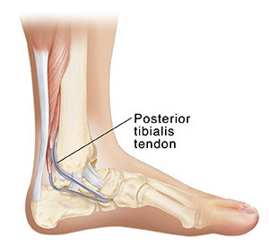
Posterior tibialis tendonitis occurs when the posterior tibialis tendon becomes overloaded and irritated. The posterior tibialis tendon starts in the calf muscle, winds under the inside ankle bone and inserts into the arch of the foot. The posterior tibialis tendon has a few key function: it helps maintain the arch of the foot, it helps invert the ankle (rotate the foot in) and it helps plantarflex the ankle (or point your toes).
(Typical) Symptoms:
The hallmark sign of posterior tibialis tendonitis is localized pain along the inside of the foot and ankle, sometimes stretching up a few inches into the shin. This might be accompanied by clicking or creaking felt within the tendon during movement as well as mild swelling along the length of the tendon. As the injury shifts from tendonitis to tendinosis the tendon becomes overloaded causing the tendon structure change and it becomes less efficient at managing loads. The tendon may not be able to provide enough stability and support for the arch of the foot, resulting in a flatting of the arch. As the condition progresses you may notice weakness when raising your heels off the ground.
Potential Causes & Their Treatments:
Initially aim to calm down and settle symptoms by managing the load placed on the posterior tibialis tendon (i.e. resting), and through treatments such as calf stretching, taping, icing, anti-inflammatory medication (if physician approved) and massage. Then identify and treat the potential cause(s). Anything that increase the load on the tibialis posterior tendon above the level that it can tolerate can be a contributing factor to the injury, such as:
Overuse - Any change in your work environment or athletic regimen - such as an increase in distance, speed or a change in terrain. It is important to allow your tissues to adapt to the increase in load. A change in job demands or training program can alter the stress placed on the tissue, and if the issue can't adapt injury occurs. Because of the progressive nature of posterior tibialis tendonitis rest is important to limit the load placed on the already injured tissue. The posterior tibial tendon is slow to heal because the portion of the tendon which runs along the inside ankle bone has a poor blood supply, limiting the rate of tissue repair.
Biomechanics - Flat feet or a tendency to overpronate places greater load on the tibialis posterior tendon. As you put weight on your foot and your arch pronates or collapses your tibialis posterior tendon plays a critical role in stabilizing your foot and maintaining your arch height. If the tendon cannot provide adequate support your arch height drops and your foot becomes more pronated, increasing tension placed on the tibialis posterior tendon, creating a vicious cycle. A shoe that provides support and has a higher heel-to-toe drop should reduce the load placed on the posterior tibialis tendon. Custom-made foot orthotics designed to improve foot and ankle position and to provide extra arch support can reduce the demands placed on the posterior tibial tendon helping to promote healing and prevent further irritation.
Muscle weakness - the main muscles responsible from plantar flexing your ankle or pointing your toes are the gastrocnemius and soleus muscles in the calf. If these muscles are weak it can place extra load on the tibialis posterior muscle because it also assists in plantar flexing the ankle. Eccentric exercises such as slowly lowering your heels down from a calf raise, resistance band exercises and toe walking can all be beneficial exercises at strengthening these muscles and tendons.
Reduced flexibility - tight calf muscles can also increase the load on the tibialis posterior tendon. Specifically calf stretches can be effective at providing relief. Hold a standing calf stretch against a wall, both with a straight knee and a bent knee, for 3 x 30-seconds twice a day.
Trauma - such as a fall or ankle sprain.
Seek out treatment from a variety of practitioners; a multidisciplinary approach can be effective at addressing the injury from multiple angles.


Lovely blog you have